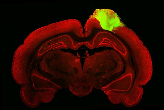
A scientific team at the University of Pennsylvania (UPenn), led by neurosurgeon Isaac Chen, has shown that brain organoids — groups of neurons grown in the laboratory — can integrate into rat brains and respond to visual stimuli, such as flashing lights. The results of the work are presented this week in the magazine Stem Cell Cell.
Previous studies have shown that individual human neurons can be integrated into rodent brains, and more recently it has been shown that human brain organoids can be integrated into rodent brains. transplanted into newborn mice. However, whether these grafts can be functionally integrated into the visual system of injured adult brains remains to be explored.
Instead of studying organoid transplantation in the intact early postnatal brain, as had been done in previous studies, we performed it in the brain of an adult rat.
Isaac Chen, neurosurgeon and leader of the work
Now, Chen explains to SINC, “Instead of studying organoid transplantation in the intact early postnatal brain, we did it in that of an adult mouse, in which a visual lesion cavity formed by aspiration. This is a fundamentally different model that focuses on brain injury and repair strategies.”
“We also examined the visual cortex —accentuates the researcher—, because it has multiple levels of neural responses that allow us to determine the degree of integration of the organoids with the host brain. In addition to responses to flashing light, we also observed that a subset of organoid neurons responded to specific directions of light stimulation. This function is called ‘orientation selectivity’ and is a unique and higher-order feature of the visual cortex.”
An architecture that resembles the brain
The team focused not only on transplanting individual cells, but also tissue. “Cerebral organoids have an architecture that resembles the brain. Thus, we were able to observe individual neurons within this structure to better understand the integration of the transplanted organoids,” says Chen.
The researchers grew neurons derived from human stem cells in the laboratory for about 80 days before grafting them into the brains of adult mice that suffered damage to the visual cortex.
After three months, the grafted organoids integrated into the rodent’s brain: they became vascularized, grew in size and number, sent neural projections, and formed synapses with the host’s neurons.
We saw that a good number of neurons within the organoid responded to specific light orientations, which shows that they were able to adopt very specific functions from the visual cortex.
Isaac Chen
The team used fluorescently tagged viruses that jumped across synapses, from neuron to neuron, to detect and trace the physical connections between the organoid and host mouse brain cells. “By injecting one of these viral markers into the animal’s eye, we were able to trace the neural connections starting from the retina”, explains the researcher, “and the tracer was able to reach the organoid”.
The image shows a neuronal marker in red, a nuclear marker in blue, and the human-specific antibody STEM121 in green, showing a transplanted organoid and its projections. / Jgamadze et al
Next, the authors used electrode probes to measure organoid neuron activity while the mice were exposed to flashing lights and alternating black and white bars. “We saw that a large number of neurons responded to specific light orientations, which shows that these organoids were capable not only of integrating into the visual system, but also of adopting very specific functions of the visual cortex”, comments Chen.
Surprising degree of integration in three months
The team was impressed with the level of incorporation of the grafts in just three months. “We did not expect to see this degree of functional integration so soon”, says the leader of the work, “there have been other studies on single cell transplantation that show that even 9 or 10 months after transplanting human neurons into a rodent, they are still not fully ripe.”
Neural tissues may have the potential to reconstruct injured brain areas. We haven’t quite figured it out yet, but this is a very solid first step.
Isaac Chen
“Neural tissue may have the potential to rebuild injured brain areas,” says Chen, who adds, “We haven’t figured it all out yet, but this is a very solid first step. Now we want to understand how organoids can be used in other areas of the cortex, not just the visual, and understand the rules that guide how organoid neurons integrate into the brain so that we can better control this process and make it work. . happen faster.”
Chen acknowledges that this research is still very experimental and that the expectations are still long term. “I think that improvements in the cellular composition and structure of current versions of organoids, as well as a better understanding of the integration process, are needed before we can think of an actual translation for patients. We are at the beginning of the path, but I am very optimistic about the potential of this strategy ”, she concludes.
Reference:
Jgamadze et al. “Structural and functional integration of human forebrain organoids with the visual system of injured adult rats”. Stem Cell Cell2023
